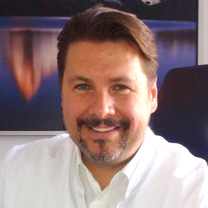
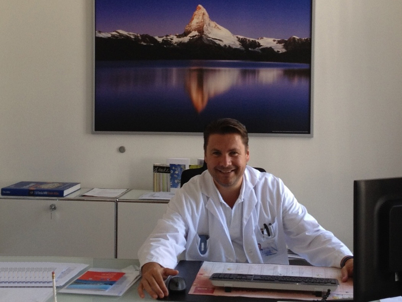
Neurosurgery Hirslanden Zurich (Switzerland): Prof. Dr Ralf A. Kockro
Treatment focus
- Microsurgery procedures for tumours of the brain, of the base of the skull and of the spinal cord
- Microvascular neurosurgery for aneurysms, cavernomata and malformations (vascular malformation) with intraoperative angiography
- Minimally invasive surgery for intervertebral disc prolapse and constricted spinal canal
- Trauma to the brain and skull with cranial reconstruction
- Microscopic and endoscopic surgery on the pituitary gland (hypophysis) and base of the skull
- Endoscopic procedures for dysfunctions of the cerebrospinal fluid circulation and tumours / cysts in the region of the ventricular system
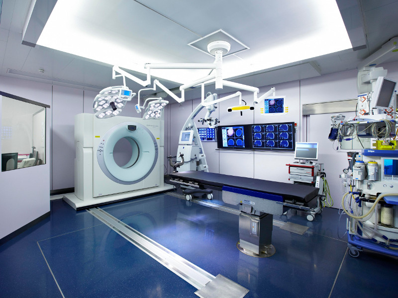
- Minimally invasive surgical planning on the 3-D simulator and surgery in open MRI and CT
Contact
Hirslanden Neurosurgery, Zurich
Center for Microneurosurgery
Witellikerstrasse 40, CH-8032 Zurich
P: +41 44 504 75 67 F: +41 44 387 21 31

Medical Range
Range of Diagnostic Services
- High-definition MRI / CT / PET imaging for precise surgical planning, in particular, functional MRI for depicting the motor centres and the speech centre
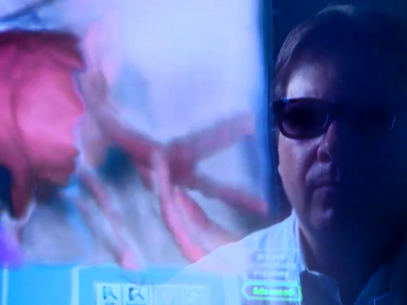
- Three-dimensional surgical planning on the virtual simulator (Dextroscope II) for preoperative simulation of the most suitable minimally invasive approach and for planning a suitable surgical strategy
- Complete specialist neurological diagnostics (MEP, SEP, AEP, NLG, VEP, EEG, Sleep Laboratory)
- Diagnosis of epileptic lesions using electrode implantation
- Diagnosis of normal pressure hydrocephalus by means of continual CSF drainage and neuropsychological assessment
Range of Therapeutic Services
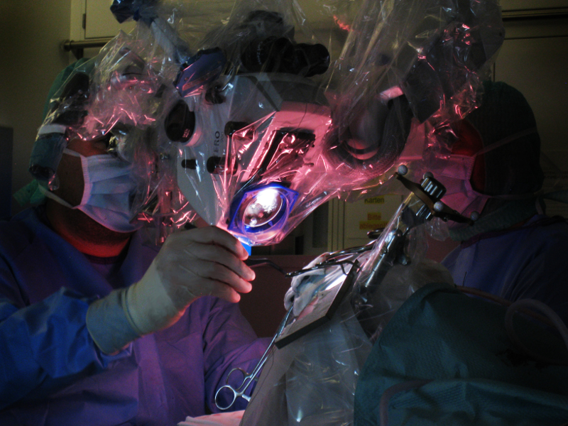
- The most up to date minimally invasive technologies in microscope- and endoscope-guided tumour surgery (e.g. glioma, astrocytoma, glioblastoma, meningeoma, neurinoma, ependymoma, metastases) with intraoperative imaging: intraoperative (open) MRI and CT, as well as intraoperative high-definition ultrasound
- Microscopic integrated fluorescence imaging of tumours using 5-ALA
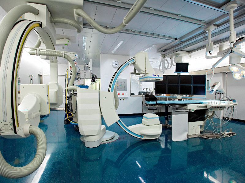
- Vascular neurosurgery (aneurysm, AVM, angioma, dural fistula), controlled by intraoperative angiography, microdoppler and ICG angiography
- Electrophysiological intraoperative neuromonitoring (MEP, SEP and cortical / subcortical mapping)
- Surgery while conscious, in particular, for procedures on the centres of the brain controlling speech and movement
- 2-D / 3-D neuronavigation including 3-D fibre tracking and fMRI
- Endoscope-assisted microvascular decompression for trigeminal neuralgia and hemifacial spasm
- Endoscope- and MRI-guided procedures for pituitary tumours (adenomata)
- Endoscopic procedures in the ventricular system for hydrocephalus, tumours and cysts
- VP shunt procedures (abdominal, endoscopic) for hydrocephalus
- Epilepsy surgery including amygdalo-hippocampectomy with electrophysiological monitoring and intraoperative EEG
- Injuries to the cranium and brain (traumatic brain injury), including the excision of haemorrhages, plastic closure, closure of CSF fistulae and cranial reconstruction with tailor-made plastic implants
- Paediatric neurosurgery for tumours, malformations and hydrocephalus
- Minimally invasive spinal surgery: microsurgical and endoscope-guided disc surgery and tailor-made minimally invasive surgery for narrowing of the spinal canal (spinal canal stenosis). Intervertebral disc replacement using cages or prostheses.
- The most up to date microsurgery for tumours of the spinal cord and tumours of the spinal canal (astrocytomata, ependymomata, neurinomata, meningeomata, pilocytic astrocytomata), including ultrasound and permanent electrophysiological monitoring
More Information
Card
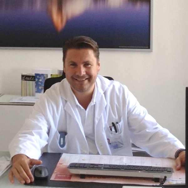
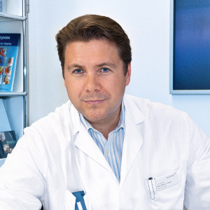
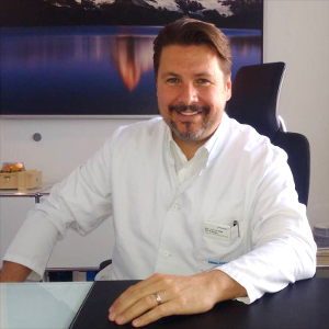
Prof. Dr med. Ralf Kockro is specialized in neurosurgery at the Hirslanden Clinic in Zurich, where he offers the complete spectrum of neurosurgery in one of the most modern clinics in Switzerland. Through the cooperation with the Hirslanden Clinic, Prof. Dr Kockro combines excellent medicine, innovative science, and the latest technical equipment in medical technology. The Hirslanden Clinic is, among others, equipped with a neurosurgical surgery room allowing intraoperative imaging, which is only available at a few locations.
Using procedures such as intraoperative MRI (magnetic resonance imaging), CT (computed tomography) and angiography (vascular imaging), diseases of the brain, skull base, and spinal cord can be treated unerringly and with maximum protection of the surrounding brain structures. This makes your surgery even safer and more precise with the best chances of success.
Perfect Surgery Planning with a Virtual 3-D Simulator
The precondition for a successful surgery is equally careful planning. Especially with inaccessible and highly sensitive structures such as the brain, every step must be well considered. Prof. Dr Kockro, and the Hirslanden Clinic have a unique virtual reality surgery planning system for this purpose. In the high-resolution three-dimensional simulator, which combines all image data from MRI and CT, Prof. Dr Kockro can simulate the entire surgery in advance. Among others, this enables him to identify the best entry path and potential danger spots.
Medical Excellence - Also in Science, Research and Teaching
Prof. Dr Kockro acquired his skills and knowledge over the years in well-respected specialized clinics in Germany, Switzerland, England, and Singapore. His additional interest in modern science allowed him to participate in important research projects for neurosurgery. Particular scientific focuses include intraoperative imaging with MRI and the development of an intraoperative navigation system.
Another of Prof. Dr Kockro's passions is teaching and training young colleagues at the medical universities of Mainz and Zurich.
Special Field Microneurosurgery
Prof. Dr Kockro specializes in minimally invasive microneurosurgery. He can gently remove the brain and spinal cord tumors in particular. Less easily accessible tumors such as the proliferation of the pituitary gland (pituitary adenoma) can be removed with the help of endoscopy (mini-camera on a thin, flexible tube).
Brain Tumors
Prof. Dr Kockro has particular expertise in minimally invasive removal of brain tumors. A brain tumor can be benign or malignant and refers to tissue growth in the brain. Due to its extensive growth, even a benign tumor can cause enormous damage by compressing neighboring brain areas. A brain tumor must be removed regardless of its characteristics.
Advanced Surgical Techniques Allow Highest Precision
Prof. Dr Kockro finds the best conditions for microsurgical tumor removal at the Zurich Neurosurgery Department. The neurosurgical surgery rooms at the Hirslanden Clinic Zurich are among the best technically equipped in the world. State-of-the-art medical technology allows unprecedented precision in planning, procedure, and execution of the surgery. Advanced intraoperative imaging provides a 3D visualization of the entire surgical procedure and simultaneous monitoring of brain functions. In interventions near the speech center, the brain tumor can even be removed when the patient is awake, which allows a highly precise procedure to avoid affecting essential brain structures.
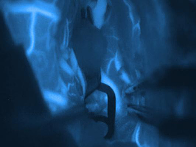
In the case of malignant brain tumors, tissue structures to be removed are marked with a fluorescent dye. The patient ingests the amino acid 5-ALA before surgery. This particular substance accumulates in tumor cells and appears bright red under blue light in the surgical microscope, which enables an exact elimination of altered tumor cells. Prof. Dr Kockro also uses endoscopes for removing tumors in some brain areas that are difficult to access, such as the base of the skull and the ventricle system.
Pituitary Adenoma
The pituitary gland is a cherry-sized hormonal gland at the base of the skull that controls important processes in the body. Tumors of the pituitary gland are usually benign and can occasionally be treated with medication. In most cases, surgical removal is required, which is carried out using the latest endoscopic technology.
Spinal Cord Tumors
If a tumor forms in the spinal cord, it compresses the spinal cord as it grows and presses on the nerve cords running in it, which leads to neurological deficits and disorders in the motor functions, which can lead to paraplegia in an advanced stage. Removal of the tumor at an early stage is always advisable.
The endoscopically removal of a spinal cord tumor is possible in the Zurich Neurosurgery Department in a microsurgical procedure. Computer-assisted monitoring accompanies the surgical procedure also to achieve the most precise process and an optimal result.
Treatments in the spinal cord are also among Prof. Dr Kockro's area of expertise. Prof. Dr Kockro is an experienced surgeon, especially in the field of herniated discs and narrowed spinal canals. Whenever possible, all interventions are carried out minimally invasive; the spectrum ranges up to the use of artificial discs (disc prostheses).
Spinal canal stenosis
The spinal cord lies well protected embedded in the spinal canal. If bony formations develop due to degenerative changes in the spinal column, they often protrude into the spinal canal. Sometimes, severe erosion of the intervertebral discs or slipped vertebrae is also responsible for a narrowing of the spinal canal. The constriction compresses the nerve structures within the spinal canal, resulting in pain, which, depending on the nerve roots affected, can spread into the legs.
If physiotherapy fails, surgical relief of the constricted nerves may be advisable. During the minimally invasive procedure, disturbing structures such as exostosis and tissue are gently removed. Sometimes the vertebral bodies have to be stabilized with the help of supporting elements to achieve the desired result.
Brain Aneurysm
Another of Prof. Dr Kockro’s specialized fields is the surgical treatment of vascular changes in the brain, which includes aneurysms and possible vascular malformations.
In the case of an aneurysm, an artery in the brain dilates to the extent that a berry or spindle-shaped bulge develops. This pathological change is usually caused by weakness of the vascular walls. Gradually, an aneurysm can grow to a size of several centimeters. Then the danger of a rupture increases. Rupture of overstrained vascular walls can lead to life-threatening bleeding in the brain. The high risk: aneurysms often cause no discomfort for a long time and are, therefore, frequently accidental findings. Depending on their position, the aneurysms can press on neighboring areas of the brain where they trigger symptoms such as impaired vision or epileptic seizures.
Depending on the shape and severity, an aneurysm can be repaired by closing the resulting bulge with a titanium clip by neurosurgery. Another option is coiling, a neuroradiological procedure, where a fine wire is forwarded through the inguinal artery into the aneurysm. Once there, it is rolled up into a ball in the bulge, which fills the entire cavity.
During the surgery, the position and treatment progress can be monitored excellently via intraoperative angiography. Prof. Dr Kockro has developed a risk calculator for the risk assessment of aneurysms. In some cases, the risk of the surgery is higher than the risk of aneurysm bleeding, so that the risk calculator can be used to prevent unnecessary surgeries. (http://www.kockro.com/)
Hydrocephalus
Close cooperation with the University of Heidelberg connects Prof. Dr Kockro to the field of brain water circulation disorders. In particular, Prof. Dr Kockro treats the various derivations of hydrocephalus, but also of cerebral fluid cysts, with the utmost care, either minimally invasive or endoscopically.
Less Stress and More Safety for the Patient
With minimally invasive neurosurgery, access to the surgery area is only as large as required, which means that this type of procedure is far less stressful for the patient than conventional surgical techniques. Besides, this type of intervention performed by experts promises an optimal result.
The possibility of virtual surgery planning should also not be underestimated. Neurosurgery Zurich is one of the few departments worldwide possessing the advanced technology of virtual reality surgery planning. This revolutionary technology allows the surgeon to run through every planned surgery from start to end and to simulate it three-dimensionally and realistically. This allows the neurosurgeon to get an exact impression of the spatial structures of a tumor or vascular changes in advance of the surgery and to plan each treatment step.
A precisely elaborated procedure and treatment concept, even before the actual surgery, not only benefits the neurosurgeon – the targeted planning also means maximum safety for the patient.
Safety has the highest priority in all of Prof. Dr Kockro’s carried out surgeries. The patient is continuously and intensively monitored during the surgery by a team of anesthetists and neurologists with the help of modern neuromonitoring (checking your nerve signals).
Click here to visit the website of Prof. Dr med. Kockro's practice.
Further information on neurosurgical topics and main focuses can be found at www.kockro.com.
Curriculum Vitae
| Since 2020 | Professor of Neurosurgery, Head of the Course Series: Minimally Invasive Neurosurgery - Virtual Reality Simulation, University Hospital Mainz |
| Since 2017 | Director of the Center for Microneurosurgery, Hirslanden Hospital, Zürich |
| Since 2012 | Senior Consultant and Co-Chair, Department of Neurosurgery, Hirslanden Hospital, Zurich |
| 2009 – 2012 | Consultant, Department of Neurosurgery, University Hospital, Zurich |
| 2008 | Consultant, Department of Neurosurgery, University Hospital, Mainz, Germany |
| 2007 | Consultant, Department of Neurosurgery, The Royal London Hospital, London, UK |
| 2006 | Senior Registrar, Department of Neurosurgery, University Hospital, Mainz; Germany |
| 1999 – 2010 | Head of the Advisory Board for Neurosurgery, Advanced Medical Technologies, Bracco AMT, Princeton, USA |
| 1999 – 2005 | Registrar, Department of Neurosurgery, National Neuroscience Institute, Singapore |
| 1999 | Co-founder of the Dextroscope Virtual Reality Technology for Minimally Invasive Neurosurgery |
| 1997 – 1998 | Fellow of the BMBF (German Ministry for Research and Technology) Department of Neurosurgery, National Neuroscience Institute, Singapore and Laboratory for Brain Imaging, National University of Singapore |
| 1995 – 1996 | Junior Registrar, Department of Neurosurgery, University Hospital Heidelberg, Germany |
| 1987 – 1994 | Medical school at the University of Heidelberg, Germany, Clinical Year at the University of California San Francisco, UCSF, Neurosurgery Department |
Extras
- Modern single and 2-bed rooms with flat-screen TV and WLAN, many with a balcony and a view of Lake Zurich
- First-class, prestigious hotels of Hirslanden Hospital
- Support for international patients by the Hirslanden International Bureau: co-ordination and organisation of consultations, medical reports, cost estimation, hotel reservations, admission to rehabilitation clinics, health spa stays, as well as transport and travel advice
- 24-hr interpreter service
- On-site underground parking
Transport Connections
| Zurich Main Railway Station | 5 km |
| Zurich airport | 15 km |
Information about Zurich
The capital of the canton of the same name is also the largest city in Switzerland. Geographically it is located north in eastern Switzerland. Zurich is situated on the Limmat River, directly at the outflow of Lake Zurich, and is thus also called the "Limmat City." Economically, socially and scientifically, Zurich is considered the center of Switzerland. Therefore, large banks, many media companies, and international companies are located there, and at the same time, the cultural offer is rich. The well-known Swiss Federal Institute of Technology Zurich (ETHZ) gives Zurich an international name in educational institutions.

