Specialists in CT (computed tomography)
6 Specialists found


Information About the Field of CT (computed tomography)
What Is Computed Tomography?
Today, computed tomography (CT) is one of the standard imaging procedures in medicine, along with magnetic resonance imaging (MRI) and sonography (ultrasound).
In contrast to the standard two-dimensional X-ray image, computed tomography is a cross-sectional imaging procedure. In this process, many transverse image slices are taken of the body or the organ in question, producing a three-dimensional image. This can be viewed and evaluated by the attending radiologist from all sides.
CT was first developed in the late 20th century (1986) by Hounsfield and Cormack. The early days were somewhat laborious, as it took about 5 minutes to acquire a single image. Today, the duration of the entire 50 individual images is only a few minutes.
The computer tomograph is a rotating tube in which a movable support table is installed. During the examination, X-rays penetrate the body from all sides. After electrical evaluation and data conversion, enable a pictorial representation of the scanned body area in grayscale.
What Are the Areas of Application for Computed Tomography?
As with any other form of imaging procedure in medicine, the benefit of CT is the acquisition of information and thereby the optimization of therapy for diseases of various kinds.
Due to its rapid performance (only a few minutes), computed tomography is usually the primary imaging for emergencies. Magnetic resonance imaging (MRI), with a 15-30 minutes duration, is inferior here. Especially in craniocerebral trauma or acute stroke, CT is one of the first measures to be taken.
Bony structures or structures with little water (bones, lungs) can be depicted particularly well in CT. In addition, areas in the upper body (thorax CT) or the pelvis (abdomen CT) can be seen particularly well.
The human body's soft tissues can generally be recognized in MRI due to better soft-tissue resolution. However, recently there is also the so-called high-resolution HR-CT (high resolution-CT) which allows particularly thin slice images (1-2mm). This makes delicate structures such as those in the lungs very clearly visible. However, HR-CT is associated with significantly higher radiation exposure.
In contrast to MRI, metal objects are not a problem in CT. Thus, even patients with metal splinters, pacemakers, etc., can be examined by CT without hesitation.
What Is the Procedure of a CT Scan?
The CT examination is a quick and painless procedure. After your attending physician has referred you to the radiologist, the examination can be started.
In contrast to the MRI, the CT is a very short tube (approx. 70 cm), so claustrophobia is extremely rare. However, if you suddenly feel uncomfortable or have problems, there is an intercom switch. You can use this to speak to the staff in the next room. You will also be monitored by camera for the entire duration of the examination.
During the examination, you will lie on a narrow couch that slowly moves into the opening of the CT. It is essential for the images to lie as still as possible. You will also have to hold your breath for a short time on command. All this is to avoid blurring of the images and prevent the CT images from being retaken (health hazard due to frequent ionizing radiation).
If the contrast between the affected organ and its surroundings is low, the radiologist can significantly increase the image quality by administering a contrast medium. The contrast medium can either be applied to the vein or, for example, drunk to examine the intestine. Particularly in the case of inflammatory processes or tumors, the contrast medium accumulates in the affected area to an exceptionally high degree, enabling the radiologist to obtain a more differentiated image and allowing more precise statements about the disease.
Once the examination is complete, the radiologist will examine the image material and hand over the findings to the attending physician, who can then adapt the therapy to the new results.
Side Effects and Risks of Computed Tomography
Computed tomography is an excellent imaging method, especially for emergencies and bony structures. The radiologist works closely with physicians of all specialties to find disease causes and guarantee the best possible therapy.
However, due to the radiation exposure, a strict indication is important because X-rays can lead to cancer formation or cell changes if used frequently. Therefore, pregnant women and children must not be examined with it or only in specific emergencies.
If a contrast medium is used in the examination, specific parameters must be clarified beforehand. Contrast media can lead to allergic reactions, including nausea, vomiting, cramps, and shortness of breath. A hormone crisis may occur in exceptional cases if a contrast medium containing iodine is used. Contrast media that are excreted through the kidneys can also cause damage to the kidneys, especially if the patient has a history of kidney disease.
If you have any further questions concerning computed tomography related to your case or risks and side effects, do not hesitate to contact the radiologist you trust.
Sources:
http://www.medizin-mit-durchblick.de/#computertomografie_anchor
Reiser, Maximilian; Kuhn, Fritz-Peter; Debus, Jürgen (2011): Radiologie. 3., vollst. überarb. u. erw. Aufl. Stuttgart: Thieme (Duale Reihe).
Kauffmann, Günter Werner; Moser, Ernst (2011): Radiologie. Bildgebende Verfahren, Strahlentherapie, Nuklearmedizin und Strahlenschutz. 4., völlig überarb. Aufl. München: Elsevier, Urban & Fischer.
Medical Articles

Your benefits
If you have found a matching specialist, you can contact him/her directly and upload records if needed. And in case you need treatment, you can…

We will direct your request to the appropriate specialists
We are happy to assist you in choosing a specialist for your needs. The service of PRIMO MEDICO is always free, confidential and discreet for…

The treatment of scoliosis in transition - When is surgery necessary?
Scoliosis is a lateral deviation of the spine. This usually involves torsion of the individual trunk sections as well as a change in the side…
![[Translate to English:] Zweitmeinung von Spezialisten](/fileadmin/user_upload/Zweitmeinung-von-Spezialisten_350px.jpeg "[Translate to English:] Zweitmeinung von Spezialisten")
Specialists' Second Opition
Many people suffer from shoulder pain or hip problems. In this case, doctors quickly recommend surgical intervention. But is this really always…
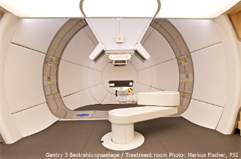
High-tech against cancer: new treatment facility put into operation
At the Paul Scherrer Institute in Switzerland a new state-of-the art treatment facility, the so-called Gantry 3, has been put into operation.
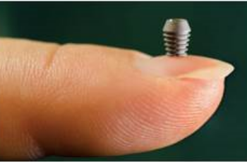
Implants: "The Longer, the Better" Has Had Its Days
Ultra-short implants have a significantly better durability than expected by experts - with lower costs, treatment times, and complications.
Modern Prostheses for Natural Walking
The ankle joint is particularly susceptible to degeneration such as osteoarthritis. It has to bear the greatest weight of all joints in the body.