Specialists in Pterygium Surgery & Radiation
4 Specialists found


Information About the Field of Pterygium Surgery & Radiation
What is Pterygium?
Pterygium is an accessory formation in the eye that originates from the conjunctiva. It usually forms in the palpebral fissure area in the inner corner of the eye. From there, pterygium grows triangularly onto the cornea and can extend far across the cornea. A gray opacity of the cornea develops in front of the tip of the pterygium. Pterygium consists of whitish translucent tissue and is interspersed by numerous newly formed blood vessels. Pterygium is a benign tumor, which frequently occurs: In Europe, two percent of people are affected by the disease! Especially in southeastern Mediterranean countries, pterygium is widespread.
What Are the Symptoms of Pterygium?
Initially, the patients have no complaints. But even before pterygium limits the vision, it can cause problems. The uneven surface can irritate the eye and affect the tear film, causing dry eyes. As pterygium continues to grow, it creates a pull on the cornea. As a result, the cornea becomes curved (astigmatism), which changes the cornea's refractive power. Incident light rays then no longer meet in a focal point on the retina but in "focal lines." As a result, the patient's vision becomes blurred. If pterygium progresses further and grows toward the center of the cornea, the patient's vision deteriorates as a result. In severe cases, when pterygium is very advanced, patients may perceive double vision. Pterygium can then even restrict the movement of the eyeball and lead to a head-restraint posture.
How Does Pterygium Develop?
It is not yet fully understood why pterygium develops. Several factors are involved in its development. However, UV light seems to play the leading role. It is assumed that hereditary factors and messenger substances (cytokines) are also involved in developing a pterygium. Dry climate and dust are also thought to advance the development of pterygium. People who are constantly exposed to a lot of UV light are more likely to develop pterygium. For example, people who live in regions with a lot of sunshine and work outdoors are particularly often affected. Glasses or darker skin color, on the other hand, protect against pterygium. It has also been found that recurrence of the disease can be prevented by consistent avoidance of UV light by wearing glasses that do not transmit UV rays or sunglasses.
UV light is thought to trigger several processes in the conjunctiva tissues: It is said to cause increased release of pro-inflammatory and vasopressor messenger substances. As a result, blood vessels and cells grow and multiply. UV light also inhibits tumor suppressor genes. Tumor suppressor genes are genes that suppress tumor growth. When they are inhibited, cells of the tissue and vessels of the conjunctiva proliferate. It is also speculated that many UV light damages stem cells in the corner of the eye in the long run.
How is Pterygium Diagnosed?
Pterygium has a typical appearance, but there are growths on the eye that resemble it. Therefore, a correct diagnosis is important. Pinguecula changes the conjunctiva that looks similar to a pterygium but does not grow onto the cornea. Malignant tumors, squamous cell carcinoma, or conjunctival intraepithelial neoplasia can also occur in the exact location. Clinically, they are sometimes hard to distinguish from a pterygium. Therefore, the physician needs to have a tissue sample examined in the laboratory after the surgery. In this way, pterygium can be distinguished from a malignant tumor. A pseudo-pterygium is a change that resembles a pterygium but is caused by an injury. However, the physician usually finds out whether an injury is a possible cause when questioning the patient.
When Does Pterygium Needs Treatment?
Pterygium needs surgery when it distorts the cornea or threatens to grow forward into the center of the cornea and limits vision. Surgical removal should be carried out in time before the pterygium reaches the cornea's center because vision often remains impaired in such cases even after surgery. Sometimes even very small pterygiums can exert a strong pull on the cornea and distort the cornea. Even then, the pterygium must be operated on. Surgery may be considered if the pterygium causes discomfort and a dry eye due to its surface. If the patient wishes, it can also be removed for aesthetic reasons.
How Is Pterygium Treated?
Pterygium is treated by surgical removal, usually in combination with medication or radiation therapy. The main problem in treating pterygium is that it often relapses after surgery. In addition, certain drugs or radiation can inhibit regrowth. Therefore, medication or radiation is usually given in addition to surgery. Several surgical techniques are combined with different drugs or radiation. However, there is no unanimous opinion among experts as to which is the optimal therapy. In the following, frequently used surgical techniques and drugs are pointed out:
- With the bare sclera technique, the surgeon excises the pterygium. The surgical removal creates a superficial wound on the sclera. The sclera is the white part of the eye, and it is covered by a thin mucous membrane called the conjunctiva. The wound on the sclera is left open and heals on its own. The conjunctiva renews itself, and the defect eventually closes. Unfortunately, with this technique, it is widespread for a pterygium to relapse.
- A widely used method is surgical removal with free conjunctival graft. After the surgeon removes the pterygium, the defect is not left open but covered with a conjunctival graft. The conjunctiva not only protects the sclera but also lines the inside of the eyelid. Next, the ophthalmologist removes the conjunctiva from the same eye under the upper eyelid. He uses it to cover the defect caused by cutting out the pterygium. The graft is sutured or glued on. Even more significant defects can be covered in this way without any problems. With this method, it is less common for the pterygium to relapse, although clinical studies have come to different conclusions in this case as well. The lasting success of the surgery depends on the size of the pterygium and the method of removal. If it was removed generously, pterygium is less likely to form a second time.
- Another method is surgical removal with an amniotic graft. After surgical removal, the surgeon covers the defect with amniotic tissue. The amniotic tissue promotes wound healing and conjunctival renewal. Again, data on the frequency with which pterygium recurs after surgery varies. However, it is higher than in the method with conjunctival graft. It is believed that the wound-healing properties may also support the regrowth of the pterygium.
- Anti-inflammatory eye drops with corticosteroids or cyclosporine are very commonly used as supportive therapy after surgery. They can be given for several weeks to months and inhibit the growth of pterygium.
- Mitomycin C is an antibiotic and also inhibits cell growth or division. It is given directly to the sclera during surgery and may additionally be given as eye drops after surgery. Mitomycin C inhibits cell division of tissue cells (fibroblasts), thus inhibiting regrowth. However, Mitomycin C has many side effects. Especially if used several times, it can cause serious consequences, such as melting of the sclera, cataract, or inflammation of the inner part of the eye. The side effects depend on the dosage. Because of the high side effects, it is recommended to give Mitomycin C only during surgery.
- Another approach is to inhibit new blood vessel formation with the angiogenesis inhibitor Bevazicumab. Pterygium is supplied by numerous newly formed blood vessels. Therefore, by inhibiting the formation of blood vessels, the pterygium should also be prevented from growing.
- Instead of drug therapy, radiation therapy with Strontium-90 may also be given after surgery to prevent a pterygium from growing again. The ß-rays inhibit cell division of rapidly growing cells. Side effects may include thinning or melting of the sclera, infection, or cataracts. However, these side effects occur very rarely. Usually, radiation is used in addition to surgery as adjuvant therapy to prevent the recurrence of pterygium. Occasionally, physicians use radiation as the sole therapy. However, this has not yet become accepted as the standard treatment.
Numerous studies have compared the various treatment methods. It was found that combined treatment showed better results in terms of pterygium recurrence than surgery without supportive treatment. Surgery with conjunctival graft achieved better results than the other surgical methods, both as sole therapy and in combination with adjunctive therapies. However, surgical methods without a graft still have their place in some cases, especially for more minor defects.
Which Doctors and Clinics Are Specialized?
The patient should look for an ophthalmologist specializing in pterygium surgery and inquire in advance which method will be used. The ophthalmologist should inform the patient in detail about the surgical method and the risks. The patient must also be informed about the side effects of the concomitant therapy. Since there is disagreement, even among experts, about the optimal treatment of pterygium, it is more difficult for the patient to decide which method and which doctor are suitable. In any case, it is recommended to look for an experienced specialist who performs this kind of surgery regularly.
We will help you find an expert for your condition. All listed doctors and clinics have been reviewed by us for their outstanding specialization in the field of pterygium and are awaiting your inquiry or request for treatment.
Literature:
- Ali. A.M. er al. (2011) The role of radiotherapy in the treatment of pterygium: a review of the literature including more than 6000 treated lesions. Cancer Radiotherapie. 2011, April;15(2):140-7
- Aminlari A. (2010) Management of Pterygium . EyeNetMagazine November/Drzember 2010. S.37/38
- Eisenmann, K. et al., Universitätsklinikum Regensburg (2020). Ergebnisse der Pterygiumchirurgie nach verschiedenen Operationstechniken – Ist die Exzision mit einfachem Bindehautverschluss noch lege artis? Der Ophthalmologe 2020, 117:359–365
- Fonseca, E., Rocha E. M., Arruda, G.V. (2018). Comparison among adjuvant treatments for primary pterygium: a network meta-analysis. British Journal of Ophthalmology. 2018, Juni, 102(6):748-756
- Grehn, F. (2012). Augenheilkunde. 31. überarbeitete Auflage, Springer Verlag
- Heindl, L.M., Cursiefen C., Augenklinik mit Poliklinik, Universität Erlangen-Nürnberg (2010). Pterygium Ätiologie, Klinik und neue adjuvante Therapien. Der Ophthalmologe 2010, 107:517–524
- Vastardis, I., (2009) Prospective Study of Exclusive Strontium-/Yttrium-90 β-Irradiation of Primary and Recurrent Pterygia with No Prior Surgical Excision Clinical Outcome of Long-Term Follow-Up. Strahlentherapie und Onkologie, 2009, No. 12, 808-814
Medical Articles

Your benefits
If you have found a matching specialist, you can contact him/her directly and upload records if needed. And in case you need treatment, you can…

We will direct your request to the appropriate specialists
We are happy to assist you in choosing a specialist for your needs. The service of PRIMO MEDICO is always free, confidential and discreet for…

The treatment of scoliosis in transition - When is surgery necessary?
Scoliosis is a lateral deviation of the spine. This usually involves torsion of the individual trunk sections as well as a change in the side…
![[Translate to English:] Zweitmeinung von Spezialisten](/fileadmin/user_upload/Zweitmeinung-von-Spezialisten_350px.jpeg "[Translate to English:] Zweitmeinung von Spezialisten")
Specialists' Second Opition
Many people suffer from shoulder pain or hip problems. In this case, doctors quickly recommend surgical intervention. But is this really always…
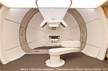
High-tech against cancer: new treatment facility put into operation
At the Paul Scherrer Institute in Switzerland a new state-of-the art treatment facility, the so-called Gantry 3, has been put into operation.
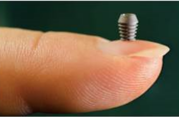
Implants: "The Longer, the Better" Has Had Its Days
Ultra-short implants have a significantly better durability than expected by experts - with lower costs, treatment times, and complications.
Modern Prostheses for Natural Walking
The ankle joint is particularly susceptible to degeneration such as osteoarthritis. It has to bear the greatest weight of all joints in the body.