Specialists in Congenital Glaucoma
6 Specialists found


Information About the Field of Congenital Glaucoma
What Is Infantile Glaucoma?
Congenital glaucoma develops after birth - usually in the first year of life. However, sometimes it is present at birth. In primary congenital glaucoma, the eye's aqueous humor cannot drain properly due to maldevelopment of the outflow tract. As a result, the aqueous humor accumulates in the eye chambers, and the intraocular pressure often increases sharply right after birth. The high pressure damages the optic nerve fibers of the retina.
The sclera, the outer shell of the eye, is even softer in young children than in adults. The increased intraocular pressure stretches the sclera and cornea, causing the eye to enlarge. This enlargement of the eye is called buphthalmos. Children are often noticed for having "big eyes." Other signs of congenital glaucoma may include photophobia, eyelid spasm, watery eyes, and a cloudy cornea. The cause of the cloudiness is corneal edema, as fluid is deposited in the cornea due to increased pressure.
Besides primary congenital glaucoma, there is also secondary congenital glaucoma, which develops due to another eye disease.
Infantile glaucoma is rare. It occurs in one in 10,000 - 18,000 children. Mostly both eyes are affected by the disease but in different degrees. If the glaucoma is not treated, the children go blind. However, the disease can be managed well with surgery in most cases.
What Is the Cause of Congenital Glaucoma?
Primary glaucoma is caused by a developmental disorder of the outflow pathways of the aqueous humor during the seventh and eighth months of pregnancy. The eye chambers are filled with aqueous humor. The ciliary bodies of the eye produce the aqueous humor. It drains away again in the chamber angle via the trabecular meshwork and the Schlemm's canal, creating constant circulation.
In most cases, the outflow of aqueous humor is obstructed by a malformation of the trabecular meshwork. More rarely, Schlemm's canal is affected by the developmental defect. Primary glaucoma occurs spontaneously or is hereditary. The cause of the defective development of the outflow tract is mutations in various genes.
Secondary infantile glaucoma can occur, for alterations, or in Peters' anomaly, a malformation of the eye. Secondary infantile glaucoma can also occur after cataract surgery.
How Is Glaucoma Diagnosed in Children?
The ophthalmologist first looks at the eye with a hand-held slit lamp during the examination. Initial indications of childhood glaucoma Enlargement of the cornea and corneal opacity may be signs of childhood glaucoma. Cracks in the posterior limiting lamina pathognomonic.
Anesthesia is required for further examination and accurate diagnosis. To determine the size of the eye, the ophthalmologist measures the corneal diameter with a caliper and the length of the eyeball with ultrasound.
The doctor can assess the chamber angle during gonioscopy, where a contact lens is placed on the eye. Through the contact glass, the ophthalmologist can see into the chamber angle. The doctor also looks at the back of the eye to see changes in the retina and optic nerve papilla. The optic nerve papilla is where the fibers of the retinal nerves bundle together to form the optic nerve, which leads from the eyeball to the brain. Hollowing of the optic nerve papilla is typical of congenital glaucoma.
Another important part of the examination is the measurement of intraocular pressure. It is 8-18 mmHg in healthy children, which is lower than adults. However, accurate determination of intraocular pressure is not easy in young children. In an awake child who is restless or crying, intraocular pressure values are often measured too high. But anesthesia can also change the values.
How Is Glaucoma in Children Treated?
Primary glaucoma in children requires surgery. However, medications are not sufficient and not effective in the long run.
Most often, a probe trabeculoctomy is carried out. In this surgical method, the doctor inserts a probe into Schlemm's canal through a small incision in the white sclera. Schlemm's canal runs around the transparent cornea in the sclera. Next, the doctor rotates the probe in Schlemm's canal. In doing so, he tears the Schlemm canal on the inside and the tissue of the trabecular meshwork over about one-third of its length. In this way, he creates a connection between the anterior chamber and the Schlemm's canal and the aqueous humor can drain away again. If this is insufficient to lower the pressure, surgery must be performed at a second site.
A new surgical method is the 360° trabeculoctomy. The doctor pushes a catheter through the entire Schlemm's canal in this procedure. Then, by pulling the ends of the catheter together, the Schlemm's canal and the trabecular meshwork are opened inward toward the eye's anterior chamber over their entire length.
For secondary infantile glaucoma, other treatment options may be considered depending on the findings, such as medications for glaucoma following cataract surgery or glaucoma drainage implants.
What Is the Prognosis for Infantile Glaucoma?
Without treatment, primary infantile glaucoma leads to blindness. However, the prognosis can be significantly improved with surgery: In 80 to 90 percent of cases, surgery is successful, and eye pressure normalizes with treatment.
The healthy eye can be covered after the surgery to avoid a loss of vision in the diseased eye, similarly to strabismus treatment, thereby exercising the other eye.
Which Doctors and Clinics are Specialized in Congenital Glaucoma?
Every patient who needs a doctor wants the best medical care. Therefore, the patient is wondering where to find the best clinic. As this question cannot be answered objectively and a reliable doctor would never claim to be the best one, we can only rely on the doctor's experience.
We will help you find an expert for your condition. All listed doctors and clinics have been reviewed by us for their outstanding specialization in congenital glaucoma and are awaiting your inquiry or request for treatment.
Sources Reference:
- F. Grehn (2012). Augenheilkunde. 31. überarbeitete Auflage, Springer Verlag 2012
- Hoffmann E.M. (2020). 360°-Trabekulotomie beim kindlichen Glaukom. Der Ophtalmologe 2020, 117:210-214
- Tamm E.R. (2011). Entwicklung des Kammerwinkels und kongenitales Glaukom. Der Ophtalmologe 2011, 108:610-617
Medical Articles

Your benefits
If you have found a matching specialist, you can contact him/her directly and upload records if needed. And in case you need treatment, you can…

We will direct your request to the appropriate specialists
We are happy to assist you in choosing a specialist for your needs. The service of PRIMO MEDICO is always free, confidential and discreet for…

The treatment of scoliosis in transition - When is surgery necessary?
Scoliosis is a lateral deviation of the spine. This usually involves torsion of the individual trunk sections as well as a change in the side…
![[Translate to English:] Zweitmeinung von Spezialisten](/fileadmin/user_upload/Zweitmeinung-von-Spezialisten_350px.jpeg "[Translate to English:] Zweitmeinung von Spezialisten")
Specialists' Second Opition
Many people suffer from shoulder pain or hip problems. In this case, doctors quickly recommend surgical intervention. But is this really always…
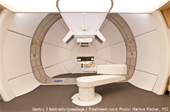
High-tech against cancer: new treatment facility put into operation
At the Paul Scherrer Institute in Switzerland a new state-of-the art treatment facility, the so-called Gantry 3, has been put into operation.
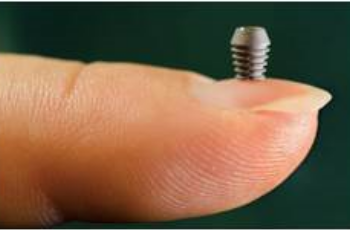
Implants: "The Longer, the Better" Has Had Its Days
Ultra-short implants have a significantly better durability than expected by experts - with lower costs, treatment times, and complications.
Modern Prostheses for Natural Walking
The ankle joint is particularly susceptible to degeneration such as osteoarthritis. It has to bear the greatest weight of all joints in the body.